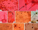
Foliar micromorphological character studies on Trichosanthes L. (Cucurbitaceae) from Terai & Duars, West Bengal, India.
Abstract
Keywords
Full Text:
PDFReferences
Abdulrahaman A.A., R.A. Oyedotun & F.A. Oladele. “Diagnostic significance of leaf epidermal features in the family Cucurbitaceae”. Insight Botany. 1(2011): 22-27. Print
Adebooye O.C., M. Hunsche, G. Noga & C. Lankes. “Morphology and density of trichomes and stomata of Trichosanthes cucumerina (Cucurbitaceae) as affected by leaf age and salinity”. Turk J. Bot. 36(2012): 328 – 335.
Ali M.A. & M.A. Fahad. “Taxonomic significance of trichomes micro morphology in Cucurbits”. Saudi J. of Biol. Sci. 18.1(2011): 87 – 92.
Bibi J.O. & B.E. Okoli. “Morphological, anatomical and cytological studies on Lagenaria breviflora (Benth.) Roberty (Cucurbitaceae)”. International J. Life Sciences. 3.3(2014): 131-142.
Chakravarty H.L. “Studies on Indian Cucurbitaceae with special remarks on distribution and uses of economic species”.(1946); Govt. of India Press: Calcutta [Reprint of the article appearing in the Indian Journal of Agricultural Science, Vol. XVI, Part I].
Chauhan D. & M. Daniel. “Foliar Micromorphological Studies on Some Members of the Family Fabaceae”. International J. of Phr and BioSci. 2.4(2011): 603-611.
Creedy J.A. “Laboratory Manual for Schools and Colleges”. Heinemann Educational Books Ltd. (1977).
Dilcher D.L. “Approaches to the identification of angiosperm leaf remains”. Bot. Rev. 40(1974):1-157.
Esau K. “Anatomy of Seed Plants”.(1959); 2 – 324. John Wiley & Sons, Inc. U.S.A.
Ibrahim G. “Microscopical studies on the leaves of Momordica charantia”. Niger J Nat Prod Med. 7(2003): 44-45.
Inamdar J.A., M. Gangadhara & K.N. Shenoy. “Structure ontogeny organographic distribution and taxonomic significance of trichomes and stomata in the Cucurbitaceae In: Biology and Utilization of the Cucurbitaceae”, eds. Bates DM, Robinson RW and Jeffrey C, Cornell University Press, Ithaca, New York.(1990):209-224.
Inamdar J.A. & M. Gangadhara. “Structure, ontogeny classification and organographic distribution of trichome in some Cucurbitaceae”. Feddes Report. 86(2008): 307-320.
Kolb D. & M. Muller. “Light, conventional and environment scanning electron microscopy of the trichomes of Cucurbita pepo subsp. Pepo var. styriaca and histochemistry of glandular secretory products”. Ann. of Botany. 94.4(2004): 515-526.
Metcalfe C.R. “Anatomy of the Dicotyledons”. Oxford Clarendon Press, U.K. 2(1950): 965-978.
Metcalfe C.R. “Current development in systematic plant anatomy”. In: Heywood V. H (Ed.) Modern methods in plant taxonomy. Academic Press, London, New York. (1968): 45-57.
Mitra S., S.K. Mukherjee & S. Bandopadhyay. Cucurbitaceae of West Bengal – a census in International Seminar on "Multidisciplinary Approaches in Angiosperm Systematics. (2005): 186-205.
Obiremi E.O. & F.A. Oladele. “Water conserving stomatal system in selected Citrus species”. S. Afr. J. Bot. 67(2001): 258-260.
Philips E.A. Method of vegetation study. (1959), Henry Holt and Co. Inc., New York, USA,107.
Rai M., S. Pandey & S. Kumar. Cucurbit research in India: a retrospect. Proceedings of the IXth EUCARPIA meeting on genetics and breeding of cucurbitaceae (Pitrat M, ed), (2008). INRA, Avignon (France).
Sharma G.K & D.B. Dunn. “Environmental modification of leaf surface traits in Datura stramonium”. Can. J. Bot. 47(1969): 1211- 1216.
Stace C.A. “The significance of the leaf epidermis in the taxonomy of the Combretaceae: conclusions”. Bot. J. L. Society. 81.4(1980): 327-339.
The Plant database. http://www.theplantlist.org. Accessed: 25th September 2018.
DOI: https://doi.org/10.21746/aps.2018.7.10.2
Copyright (c) 2018 Annals of Plant Sciences

This work is licensed under a Creative Commons Attribution-NonCommercial-NoDerivatives 4.0 International License.


